For about two years, specialists from the Republican Scientific and Practical Center for Traumatology and Orthopedics have been cooperating with the High-Tech Park of Belarus in the field of implementing innovative methods of treating patients. In 2019, a joint project of the Center and Innowise Group in the field of augmented reality Voka.io won the international World Summit Awards in the Health and Well-being category (held with the support of the UN). The development was presented at a congress of traumatologists and orthopedists in the USA that same year, and in 2018 - at a European congress of traumatologists, where the project also won prizes. The essence of the idea is to use mixed reality in traumatology. IT specialists from Innowise Group and Alexander Sitnik, head of the laboratory of adult traumatology at the Republican Scientific and Practical Center for Traumatology and Orthopedics, are working on the unique Voka.io project.

How is modern medicine changing under the influence of technology, how will people be treated and is it possible to dream of a future without diseases today? The "Clinics of Belarus" spoke about this with traumatologist-orthopedist Alexander Sitnik.
What are the main objectives of the project implementation?
In this project, each of its participants faces their own professional challenges. For the doctor, this is the practical application of innovation, and for IT specialists, this is the technological implementation of the plan. In simple terms, in addition to routine situations, there are situations when doctors face complexities of both anatomy and fracture morphology, which require maximum, special precision. Given the specifics of our activities, we decided to use complex, intra-articular, comminuted fractures of the lower limb bones as the area of study, the peculiarity of which is that not only bones but also soft tissues suffer. And as is known, bone healing depends on soft tissues, therefore, in order to get a good result, not only a clear picture on the X-ray is necessary, but also high-quality wound healing. Therefore, it is very important to plan the operation in detail and subsequently perform surgical access in specifically designated places.
How does this work?
Let's say we receive a patient (we do not consider emergency situations) with a complex fracture of the distal tibia from a fall from a height. The injury pattern is a high-energy trauma, the bone is crushed "into splinters", soft tissues are also damaged. And so, the first stage is to apply a conventional rod apparatus for external fixation, stretch the leg, partially reposition the fragments, after which a CT scan is performed. Having completed these manipulations, we can already proceed to the second stage - direct planning of the operation. It should be noted that in the absence of a critical moment for life, several days pass between the injury and the operation. This is due to the medical process: a person is given antibiotics to, as doctors say, calm the soft tissues. During this period, preoperative planning occurs, when medical data from an MRI study is converted into a 3D model created by IT specialists, after which the fracture model of a specific person is loaded into HoloLens (AR glasses). The doctor can rotate, scale and study the fracture elements with his hands without having to touch anything on the patient's broken leg. This model allows you to understand how many key elements there are, what, so to speak, gap between them you need to get into to put the articular surface evenly and determine some key points that are required for the operation.
What is the situation with comminuted fractures, or more precisely with their small fragments?
It all depends on the threshold set, some small fragments may be lost, but they are not so important to us in life.
The operation plan is made, what happens next?
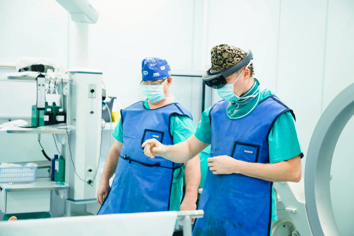
The plan is drawn up, the necessary access is determined, the fracture line is established through which the joint is reached and the surface itself is restored. The purpose of such planning is to minimize soft tissue trauma, i.e. the surgical incision should pass specifically above the planned fracture line. Accordingly, the next stage of work is already directly in the operating room. When the patient is on the operating table, the surgeon puts on AR glasses, places the model of the patient's fracture on his leg, using anatomical landmarks. Thus, the leg and the fracture are in the doctor's field of vision, after which a marker is taken and the planned surgical access is marked. Then, without using glasses, the operation itself is performed.
How many real operations have already been performed using the new technology?
Today there are about eighty of them.
In what other areas of medicine is it planned to use this technology?
The Republican Scientific and Practical Center for Pediatric Surgery conducts research with a group of pediatric cardiac surgeons, and in the field of oncology, there is also cooperation with the Republican Scientific and Practical Center for Oncology and Medical Radiology named after N.N. Alexandrov.
What are the prospects for further development of your project?
At the moment we are working with a drawn bone, in the future we expect to create models in which vessels with nerves will be displayed in accordance with data from examinations of a specific patient.
Are mixed reality glasses used in the educational process?
They are already widely used in the educational process. This is primarily the study of anatomy, where an anatomical atlas is loaded in three-dimensional resolution and the student can examine a particular organ from all sides, which significantly simplifies and shortens the learning process.
At the moment, the whole world is actively moving towards the implementation and development of innovations in the field of medicine, and Belarusian doctors, together with IT specialists, are confidently declaring themselves in the international arena.